

BIX-L1007 Review: Transparent Adult Tooth Model — Why 16 Indicators Beat a Bigger Model
Article tag: Transparent adult tooth model BIX-L1007 L1007
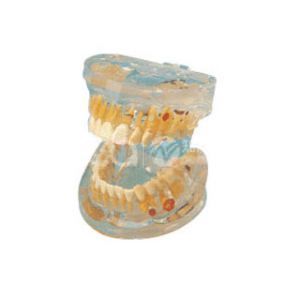
BIX-L1007 transparent adult tooth model: maxillary and mandibular parts in two sections showing the form and structure of adult teeth through 16 indicators, so students can see the full adult arch, its occlusion and its internal structure, for dental anatomy and oral hygiene teaching. ISO and CE. US $82.50. Email sophia@adahealthy.com.
...
Product Description
Model | BIX-L1007 — Transparent Adult Tooth Model |
Summary | Two-part transparent adult tooth model, maxillary and mandibular, with 16 indicators showing adult tooth form and structure for dental anatomy teaching. |
Structure | Maxillary teeth and mandibular teeth as two separate parts, with 16 indicators marking the form and structure of the adult teeth |
Transparency | Transparent material, so internal structure is read from outside rather than only the visible crown |
Certification / Price | ISO & CE (as stated); US $82.50, factory-direct with flexible MOQ |
Educational-use note: teaching model — educational equipment, not a medical device.
Before ordering: the specification PDF is not published, and the price appears on the category listing but not in the product page body. Request the datasheet: sophia@adahealthy.com.
1. What the L1007 Is
The L1007 is a two-part transparent model of the full adult dentition — maxillary teeth and mandibular teeth held as separate parts — carrying 16 indicators that mark the form and structure of the adult teeth.
The two-part build gives you occlusion. The arches separate, so an instructor can show how the maxillary and mandibular teeth meet, then lift the upper arch away to expose what the contact hides — impossible on a single fixed arch.
Transparency gives you the inside. An opaque model teaches only what the eye can reach: crown outline, cusp count, grooves. A transparent one shows the internal structure, crown to root, without sectioning anything.
In the manufacturer's transparent group: L1009 (baby teeth development, US 134.08), L1007 (adult, US 82.50).
2. The Learning Objective It Has to Serve
Dental anatomy has specific, testable objectives: identifying the anatomical and morphological characteristics of human primary and permanent teeth, identifying and reproducing tooth surface details in order to recognise and diagnose anatomical changes, and developing the psychomotor skills to restore teeth with proper form and function. Most schools still deliver that through lectures plus two-dimensional drawings of teeth, identification of features in preserved teeth, and carving. The criticism is already published: oversized carving materials may distort students' understanding of anatomical proportions, and lecture-driven instruction is limited in fostering interactive learning, producing a gap between pre-clinical instruction and practical patient care.
Read that against this model's specification. A model that is enlarged for convenience and opaque for durability reproduces exactly those two problems: the proportions are wrong, and the anatomy you cannot see is the anatomy you cannot diagnose. The L1007 answers differently — correct adult scale, and a material that shows the inside.
3. What Transparency and 3D Handling Actually Change
Rotation and spatial understanding are measurable. A cross-over study built a 3D tooth morphology quiz application that lets students select teeth, change viewpoint and zoom, and rotate the tooth to obtain a three-dimensional spatial understanding of its different surfaces. Studying with it produced an 18% increase in scores over three weeks (P < 0.001), and assessment scores were correlated with app scores while not being influenced by which teaching modality students accessed first — tooth identification is a spatial task, and turning the object in your hands is what the app simulated.
Transparency plus sectioning is the accepted design logic. When a dental school designed 3D-printed macro-models of a mandibular first molar at ×9 enlargement, it produced them on a stereolithography printer in transparent liquid resin — one showing the optimal endodontic access cavity, and a second divided along a mesio-distal axis to visualise the root canal system. Most students found these macro-models effective for endodontic training. Designers chose transparent resin and a split model for one reason: the decisive anatomy in a tooth is internal.
And the tooth-shaped form is already preferred. In a study of a 3D-printed practice tooth reconstructed from a micro-CT scan of an extracted tooth 36, students rated both the 3D-printed tooth and natural tooth models significantly better than transparent acrylic blocks — suitability as a practice option (p < 0.001) and handling (p = 0.001). The acrylic block, the incumbent in most pre-clinical labs, rates worst because it has no morphology to read. Morphology is the product; transparency only helps when there is anatomy to see through it.
4. What the Model Does Not Do
It exercises recognition, not clinical skill. A study of 99 undergraduate dental students surveyed knowledge of master gutta-percha cone selection, then observed 45 of them fitting cones on real patients. 80.8% knew how to properly manage a short cone — but the proper flaring assessment method was chosen by 86.9% of the female students against 34.2% of the male students (p = 0.0001). Naming the tooth and judging the case are separate skills — the authors' framing is that misconceptions should be detected early. A model is a recognition and discussion tool; hand skills still need clinic time.
The adult arch is not the early-years case. This is permanent dentition — L1009 / L1006 cover deciduous teeth and L1008 transparent pathology. Buying the adult arch for a paediatric unit buys the wrong specimen.
5. Line Placement and Buying Logic
Where it sits. The L1007 is the full adult arch, transparent option. Around it: L1018 (full mouth transparent soft rubber, US 50.51); L1001 (amplification, US210.47) where enlargement is wanted.
Buying logic: buy the L1007 where a course must teach identification of the permanent dentition, the maxillary-to-mandibular relationship, and what is inside a tooth without sectioning one. Where the subject is caries, periodontal disease or pulp pathology, the pathological models in the same range are the correct instruments. At US $82.50 it is a low-cost per-desk item.
Checklist (identification drill)
● Every tooth in both arches named by type and position, using the model alone
● Maxillary and mandibular arches separated, then related to show occlusion
● The 16 indicators used as a scripted route through form and structure
● Internal structure traced on the transparent model, then matched to the corresponding radiograph
● Surface details reproduced on a carving block and compared back to the model
6. FAQ
Q1: What is the BIX-L1007? A: A transparent model of the full adult dentition, built as two parts — maxillary teeth and mandibular teeth — with 16 indicators showing tooth form and structure.
Q2: Why transparent rather than opaque? A: So students see the internal structure from outside — the same choice made where transparent resin models visualise the root canal system.
Q3: Why does the two-part build matter? A: The arches separate, so the maxillary-to-mandibular relationship and occlusion can be demonstrated.
Q4: What is the price? A: US $82.50, factory-direct with flexible MOQ. Email sophia@adahealthy.com for quotation, bulk pricing and the datasheet.
Q5: Will a model alone make a student clinically competent? A: No. In one study 80.8% knew how to manage a short cone, yet technique selection still differed sharply within the cohort.
Q6: Is it suitable for baby teeth or pathology? A: No. This is the adult permanent dentition; use BIX-L1009 / L1006 for deciduous teeth and BIX-L1008 for transparent pathology.
References
Flipping the Dental Anatomy Classroom (2018)
Different Methods of Teaching and Learning Dental Morphology (2024)
A 3D Tooth Morphology Quiz for Dental Students (2019)
3D-Printed Macro-Models for Endodontic Access Cavities (2022)
3D-Printed Tooth for Endodontic Training (2025)
Master Gutta-Percha Cone Selection: Knowledge vs Performance (2024)

Marketing Center
Hong Kong, ChinaProduction Base
Shanghai, ChinaProducts
Contact Us
 Address: Hong Kong, China
 Phone:+86 13383897707
 Email:sophia@adahealthy.com
 Mobile:+86-0379-65160607
举报邮箱:sophia@adahealthy.com
举报电话:+86 19937901373
Social Media