

BIX-F9A Guide: Uterine Cervical Lesion Model — Six Cervical Lesions & Two Normal Uteri
Article tag: Uterine cervical lesion model BIX-F9A F9A
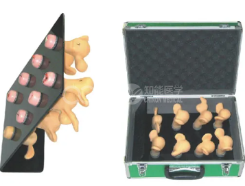
BIX-F9A uterine cervical lesion model: six cervical lesions (erosion, linear laceration, acute suppurative cervicitis, cancer, polyps, follicular cyst) and two normal uteri in one visual teaching aid for medical, health and nursing schools, for pathology recognition. ISO and CE. US $235.68. Email sophia@adahealthy.com.
...
Product Description
Model | BIX-F9A — Uterine Cervical Lesion Model (Cervical Pathology Teaching Trainer) |
Summary | Uterine cervical lesion model: six cervical lesions — erosion, laceration, suppurative cervicitis, cancer, polyps, follicular cyst — plus two normal uteri. |
Lesion Set | Six lesions: erosion; linear laceration; acute suppurative cervicitis; cancer; polyps; cervical gland (follicular) cyst — plus two normal uteri for comparison |
Intended Use | Visual teaching aid during clinical practice, for students in medical and health schools and hospital paediatrics |
Users | Physicians and health educators; nursing and midwifery teaching |
Certification / Price | ISO & CE (page-stated); US $235.68 per set |
Educational-use note: educational equipment, not a medical device for patient use. Request the spec sheet by email for spare parts and set contents.
1. Why Visual Recognition Deserves a Bench Model
Cervical pathology is learned by eye. Whether the assessment is naked-eye inspection, colposcopy or a screening programme, the job is to look at a cervix and decide: normal, or not normal. That decision is trainable — and the evidence on how well it is made explains why a lesion set belongs on the bench.
Start with scale. A meta-analysis pooled 23 studies and 101,273 women to measure the accuracy of visual inspection after acetic acid (VIA) and after Lugol's iodine (VILI). Pooled sensitivity of VILI was 88% and specificity 86%, while positive predictive values were low, ranging from 11% to 16%. The authors concluded that although imperfect, VILI alone appeared to be the most useful visual screening strategy (Catarino et al., 2018). A test that turns on an eyeball judgement deserves deliberate practice.
Second, structured criteria help measurably. In a prospective study in Cameroon, 1,980 women were screened (361, or 18.4%, HPV-positive); assessors applying the ABCD criteria — acetowhiteness, bleeding, colouring, diameter — reached a sensitivity of 77.5% (95% CI 61.3–88.2) and a negative predictive value of 93.3% for CIN2+, with 86.7% of positive women treated the same day (Petignat et al., 2022). Named, defined appearances are what make such criteria teachable.
Third, skill level shows up plainly. When 22 healthcare workers and 9 experts assessed the same 83 cervical images, specificity was 80.5% versus 93.5%, and inter-observer agreement (Fleiss kappa) was 0.45 versus 0.68; the paper notes that visual inspection is limited by subjectivity and a lack of skilled human resource (Nakisige et al., 2023). Agreement of 0.45 is not a knowledge gap; it is a recognition gap.
Maintaining that skill is also structural: although WHO recommends VIA, quality varies widely and it is difficult to maintain a well-trained cadre of providers (Ferguson et al., 2024). Physical lesion models let a trainee compare the same six appearances repeatedly, without a patient.
2. Evidence
Evidence | Finding | Relevance |
Catarino et al., 2018 | 23 studies, 101,273 women: VILI sensitivity 88%, specificity 86%; PPV low (11–16%) | Visual assessment is useful but imperfect |
Petignat et al., 2022 | 1,980 women, 361 HPV-positive: ABCD criteria gave sensitivity 77.5%, NPV 93.3% | Named features make assessment teachable |
Nakisige et al., 2023 | 22 workers vs 9 experts, 83 images: specificity 80.5% vs 93.5%; kappa 0.45 vs 0.68 | Recognition separates skill levels |
Ferguson et al., 2024 | WHO recommends VIA, but quality varies and maintaining trained providers is difficult | Rehearsal must be repeatable |
Kiesel et al., 2022 | 3D-printed LLETZ simulator: simulation lacked realistic cervical dysplasia anatomy | Model teaching closes a gap |
González Méndez et al., 2025 | 13 physicians: correct diagnoses rose from 47% to 80% (p < 0.001) | Training improves accuracy |
3. What the F9A Lets You Teach
A. Six lesions, one bench
Erosion, linear laceration, acute suppurative cervicitis, cancer, polyps and a cervical gland (follicular) cyst are presented as separate specimens, so a session moves from benign to malignant without changing rooms.
B. Normal is taught as a category
Two normal uteri are included for side-by-side comparison. The hardest judgement in visual assessment is not spotting florid disease but deciding that an appearance is within normal limits (Petignat et al., 2022).
C. Targeting the appearances that are missed
Agreement between healthcare workers and experts diverges most on specificity (80.5% vs 93.5%) rather than sensitivity (Nakisige et al., 2023), so the useful drill is not "spot the cancer" but "justify why this is not abnormal" — which the normal uteri, the cyst and the erosion specimens supply.
D. The bench step before colposcopy
Described as a visual teaching aid during clinical practice, the set is where structured terminology — acetowhiteness, bleeding, colouring, diameter — becomes a rehearsed vocabulary rather than a list (Petignat et al., 2022).
4. Where the F9A Sits in the Line
Model | Scope | Best for |
F9A | Six cervical lesions + two normal uteri | Pathology recognition |
F9 | Advanced cervical change model | Progressive cervical change |
F9B | Female contraception model | Contraception education |
F9E | IUD placement trainer | Intrauterine device insertion |
F100 | Female pelvic contraceptive model | Family-planning teaching |
F5M | Male condom practice model | Contraception education |
Buying logic: choose the F9A when the curriculum needs named lesions with normal controls; add F9E for IUD insertion, or F9 when progressive cervical change must be demonstrated over time.
5. Teaching Protocol
Station | Time | Activity |
A. Normal pattern | 10 min | Study the normal uteri; agree on normal limits |
B. Benign set | 20 min | Identify erosion, laceration, suppurative cervicitis |
C. Malignant and structural | 20 min | Examine cancer, polyps, follicular cyst; justify each call |
D. Blind round | 15 min | Unlabelled specimens; state a diagnosis and a reason |
E. Debrief | 10 min | Compare answers with the list; correct terminology |
Assessment checklist
● All six lesions named correctly without labels
● Normal specimens classified as normal
● One specific visual feature given as the reason for each answer
● Benign and malignant appearances distinguished
● Terminology used consistently
6. Maintenance
Item | Frequency | Notes |
Specimen surfaces | Each session | Mild disinfectant; no solvents |
Case and labelling | Monthly | Confirm each specimen is in its slot |
Storage | Daily | Cool, dry, out of sun; seated, not stacked |
7. FAQ
Q1: What is the BIX-F9A? A: A uterine cervical lesion model supplied as cervical pathology specimens: six lesions — erosion, linear laceration, acute suppurative cervicitis, cancer, polyps and a cervical gland (follicular) cyst — plus two normal uteri.
Q2: How many pieces are in the set? A: Six lesion specimens and two normal uteri. Request the spec sheet at sophia@adahealthy.com. for the contents list.
Q3: Who is it intended for? A: Students in medical and health schools, hospital paediatrics and clinical teaching, plus physicians and health educators.
Q4: Why include two normal uteri? A: Because the difficult judgement is deciding that an appearance is within normal limits. In an image-based study, specificity separated healthcare workers from experts far more than sensitivity did (80.5% vs 93.5%).
Q5: What is the price and MOQ? A: Listed at US $235.68 per set; MOQ 1 set. Pricing depends on configuration — email sophia@adahealthy.com. for the quotation.
Q6: What certifications and shipping terms apply? A: ISO & CE as stated on the page; air freight 7–10 business days, sea freight 30–45 days. Details: sophia@adahealthy.com..
References
Accuracy of Visual Screening for Cervical Precancer (Catarino et al., 2018)
ABCD Criteria for Visual Cervical Assessment (Petignat et al., 2022)
Reader Agreement in Cervical Image Assessment (Nakisige et al., 2023)
Maintaining Screening Quality in Practice (Ferguson et al., 2024)
3D-Printed Cervical Dysplasia Simulator (Kiesel et al., 2022)
Colposcopy Training Platform Outcomes (González Méndez et al., 2025)

Marketing Center
Hong Kong, ChinaProduction Base
Shanghai, ChinaProducts
Contact Us
 Address: Hong Kong, China
 Phone:+86 13383897707
 Email:sophia@adahealthy.com
 Mobile:+86-0379-65160607
举报邮箱:sophia@adahealthy.com
举报电话:+86 19937901373
Social Media