
How does the cephalic artery model show the cerebral artery system?
Article tag: Cephalic artery model anatomical model of the head

In summary, the cephalic appendicular artery model provides learners with an intuitive and comprehensive display platform of the cerebral artery system by accurately displaying the structure of main and branch arteries, indicating the direction of blood flow, using color and marking, and providing detachable or segmented classification functions. This not only helps medical professionals to have a deeper understanding of the structure and function of the cerebral artery system, but also appl...
When presenting the cerebral artery system, the cephalic appendicular artery model is usually used in an accurate and intuitive way, so that learners can clearly understand and grasp the structure and function of the cerebral artery. Here is a detailed explanation of how the model specifically demonstrates the cerebral arterial system:
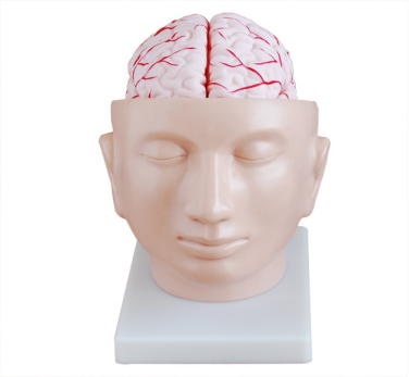
Presentation of the main artery:
The model first highlights the two main main cerebral arteries, the internal carotid artery and the vertebral artery. The internal carotid artery enters the cranial cavity through the carotid foramen of the temporal bone and forms a U-shaped structure after passing through the cavernous sinus near the sphenoid bone. This structure is a common site of arteriosclerosis. The vertebral artery supplies the posterior 1/3 of the cerebral hemisphere and parts of the diencephalon, brainstem, and cerebellum.
Detailed depiction of branch arteries:
The model further delineates the branches of the main artery, such as the middle cerebral artery, the anterior cerebral artery, and the posterior cerebral artery. The middle cerebral artery is a direct continuation of the internal carotid artery and supplies most of the lateral surface of the cerebral hemisphere. The anterior cerebral artery supplies blood to the medial side of the brain before the parieto-occipital sulci; The posterior cerebral artery runs backward along the lower part of the brain, supplying the occipital lobe, the medial side of the temporal lobe, and the basal side of the brain.
Indication of blood flow direction:
Arrows or other signs are used to indicate the direction of blood flow in the cerebral artery system to help learners understand the blood supply path in the cerebral artery system.
Use of colors and markers:
The model may use different colors to distinguish different arteries and branches, or use specific markers to label important arterial structures and anatomical points, making the entire cerebral artery system clearer and easier to understand.
Detachable or separable parts:
Some advanced models allow users to disassemble or separate partial structures in order to more deeply observe and understand the various parts of the cerebral arterial system. For example, users can remove parts of the skull to see how cerebral arteries are distributed inside the skull.
Detailed annotation and description:
The model is usually accompanied by detailed annotations and instructions, explaining the name, function, and role of each artery and branch in the blood supply to the brain, to help learners acquire a more comprehensive knowledge of the cerebral artery system.
In summary, the cephalic appendicular artery model provides learners with an intuitive and comprehensive display platform of the cerebral artery system by accurately displaying the structure of main and branch arteries, indicating the direction of blood flow, using color and marking, and providing detachable or segmented classification functions. This not only helps medical professionals to have a deeper understanding of the structure and function of the cerebral artery system, but also applies to popular science education and public understanding.
Marketing Center
Hong Kong, ChinaProduction Base
Shanghai, ChinaProducts
Contact Us
 Address: Hong Kong, China
 Phone:+86 13383897707
 Email:sophia@adahealthy.com
 Mobile:+86-0379-65160607
举报邮箱:sophia@adahealthy.com
举报电话:+86 19937901373
Social Media